of Mental Illness and Other Brain Disorders
- Addiction >
- Analysis &
Informatics > - Autism >
- Forensics >
- Neurodevelopment >
- Psychosis >
- Traumatic Brain Injury >
Addiction research at MRN focuses primarily on two main areas — the striatum (in red) and anterior cingulate (in blue). These areas activate more strongly in response to alcohol and drug cues than they do in response to other appetitive cues, such as a sugary drink.
The analysis and informatics capabilities at MRN include: advanced multivariate algorithms and software tools, graph theoretic measures, approaches for fusing multimodal brain imaging and genetics data, prediction of outcomes, automated analysis pipelines and centralized data management and storage.
more about our work with Neuroinformatics >
more about our work with Medical Image Analysis >
Primary autism research focuses on two brain circuits, one supporting auditory processing and language function (blue), and the other supporting social cognition and face processing (red). Additional research explores multimodal sensory processing, epilepsy in autism, and the development and evaluation of novel treatments.
Psychopathy research at MRN examines the overlapping contributions of substance abuse and psychopathy. Psychopathy dysfunction is in the limbic brain regions (blue), anterior and posterior cingulate cortices (blue and red) and amygdala. Substance abuse is also found in the prefrontal regions (blue), as well as the basal ganglia (green).
MRN researchers study relationships between connectivity and structure of circuits involving the anterior cingulate gyrus (red), medial orbital frontal lobe (blue) and cerebellum (green), characterizing differences between healthy children and those born prematurely and with other developmental disorders.
Schizophrenia affects communication between areas in the frontal and temporal lobes (in red). In bipolar disorder the key areas include the inferior frontal lobes and the more “emotional” parts of the brain (not shown). Parietal areas (in blue) can be affected in both psychoses.
Traumatic Brain Injury research at MRN suggest subtle lesions (green) following mild TBI, including metabolic and functional abnormalities in otherwise healthy appearing tissue. These lesions are often missed because they are not located in common sites of functional disturbances (red) and white matter injury (blue).
Inside MRN //
Headquartered in Albuquerque, New Mexico, MRN is a 501(c)3 non-profit organization, consisting of an interdisciplinary association of scientists located at universities, national laboratories and research centers around the world and is focused on imaging technology and its emergence as an integral element. read more >
Meet Our Investigators //
John Phillips, MD

Dr. Phillips is a licensed physician who, as MRN's Medical Director, has responsibility for ensuring the safety and ethical conduct of clinical and research operations.
His expertise and research are in functional neuroimaging, pediatric neurorehabilitation, cerebral palsy, spasticity management and the care of children with special needs in Eastern European orphanages.
Imaging Services & Capabilities //
Elekta Neuromag Vector View 306 Channel Meg
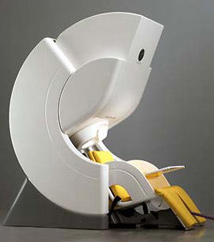
This 306 channel MEG system is comprised of 204 planar gradiometers and 102 magnetometers and is capable of collecting up to 128 channels of simultaneous EEG data at up to 5 kHz sampling frequency. The planar gradiometers provide sensitivity to sources close to the sensor array whereas the magnetometers provide sensitivity to distant sources, including sources originating from the…